

Authors
|
|||||
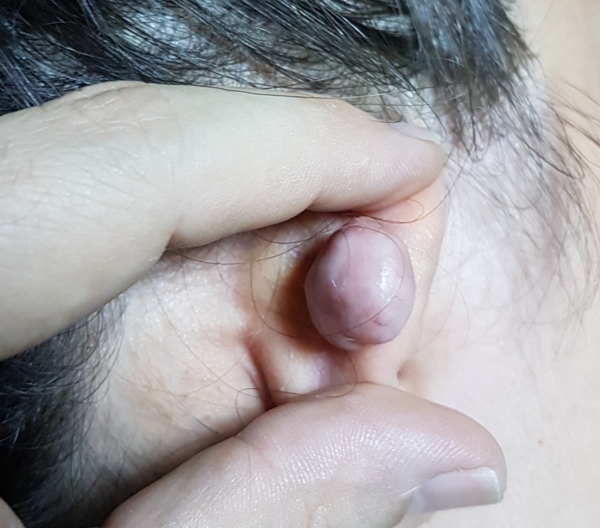
AbstractKeloids are benign proliferative lesions caused by abnormal healing. Treatment of keloids is very difficult due to high recurrence rates. In keloids, there are many treatment modalities that can vary according to lesion size, localization, and previous interventions. A 17-year-old female patient presented with a posterior ear keloid that developed after pearcing into her ear. The shaving excision was treated with a triple combination consisting of single session intralesional steroid injection and daily topical imiquimod application for six weeks. Within one year, a very good response was obtained without recurrence or any side effect.IntroductionKeloids are abnormally proliferative benign lesions caused by exaggerated healing. They create considerable cosmetic problems for the patients. There is also a potential for skin cancer development [1]. Ear is one of the localizations where keloids are frequently seen because it is preferred to wear earrings. Ear keloids are a region that creates a serious cosmetic problem as it is in a visible region and also poses a challenge for the aggressive treatment needed for keloid therapy [2]. We present a young patient with a big keloid in her pinna for two years. She was treated successfully with a combination of shave excision and intralesional steroid injection followed by topical imiquimod application. To the best of our knowledge, this triple combination has never been used in the treatment of auricular keloid in the literature. Case ReportA 17-year-old female patient admitted to the dermatology department with a massive mass complaint behind the right ear (Figure 1).
DiscussionKeloids are difficult skin lesions to treat. Uncontrolled surgical operations can lead to larger keloids. Prevention of recurrence in keloid treatment should be the main objective. Imiquimod is one of the agents used to prevent recurrence. Imiquimod is a strong inducer of interferon. The possible role of interferon is to suppress the activity of the keloidal fibroblasts that cause keloid formation [3]. Topical imiquimod has been used alone or as part of combined treatments in keloid treatment. In addition to successful results, ineffectiveness reports have also been reported [4-7]. Bermann and Kaufman used imiquimod for eight weeks immediately after surgery and did not detect recurrence [4]. There are other studies showing that use of imiquimod after surgery in the treatment of auricular keloids reduces recurrence as well as our case [5,6]. But there is also a study reporting that topical imiquimod has failed to prevent recurrences after keloid surgery [7]. But the keloids tried to be treated in this study were not at auricula but at the trunk [7]. We performed intraoperative triamcinolone injection into the shaved keloid base in addition to shaving excision and postoperative topical imiquimod treatment in our case. It is often difficult to inject triamcinolone into the keloids due to its hardness. Injecting into the lesion base immediately after excision is much easier than injecting into the keloid. Also, triamcinolone injection into the keloid is not effective without excision in large keloids. When triamcinolone injection is used, we think that the use of topical imiquimod will be sufficient for a shorter period of time than the application of the eight weekly topical imiquimod, which is frequently applied in the literature. Radiotherapy after surgical excision is an alternative method in the treatment of keloid [8]. However, an optimal protocol for total dose and fractions of adjuvant radiation for ear lobe keloids has not yet been established. Therefore, we did not consider radiotherapy in the treatment of our case. Liu and Yuan [9] suggest that three-fraction electron radiotherapy after excision within 2 days of surgery is a safe and effective protocol for the prevention of earlobe keloid recurrence that can also improve patient compliance and comfort. Ragoowansi et al. [8] applied a single dose of radiotherapy 24 hours after surgical excision. Early radiotherapy after surgical excision may be preferred, especially in keloid patients who do not benefit from previous treatments and have a high risk of recurrence. However, it has been reported that cancer develops after radiotherapy for keloid treatment, although it is very rare [10]. Conclusion As a result, we can deal with big challenging keloids using the advantages of all methods in surgical excision combined with triamcinolone injection and topical imiquimod. References
|
|||||
| Keywords : keloid , shave eksizyon , intralezyonel , steroid , imiquimod. | |||||

|