

|
|||||||||
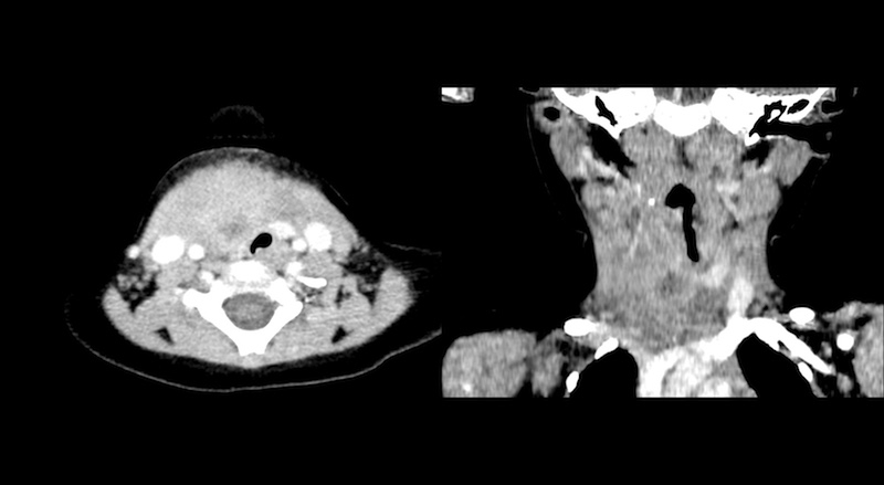
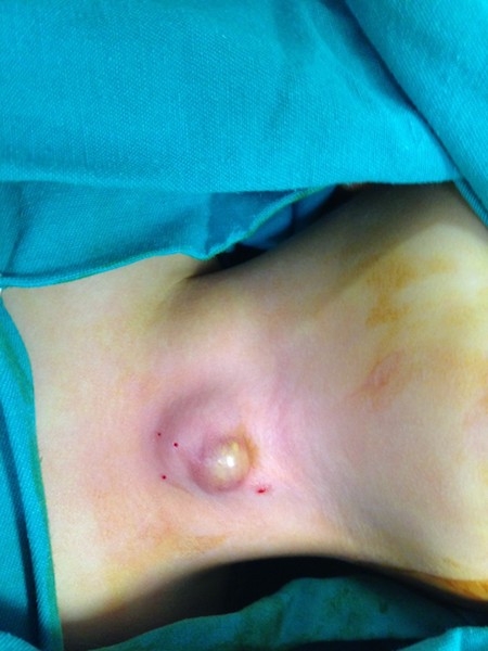
AbstractForeign body existence in oropharynx and larynx is frequently encountered in emergency department and otorhinolaryngologic outpatient clinics. Foreign objects that are ingested by children include common household items like coins, small toys, marbles, batteries, erasers. We present an unusual case of patient with neck abscess caused by a harmless foreign body migrated from larynx to the neck. Although our clinic is an experienced clinic for foreign body ingestion and aspiration, we should say that we did not confronted with this kind of case previously. We think that presenting that kind of cases into literature is important due to attract physicians attention.IntroductionForeign body ingestionis a very commonly seen problem in pediatric population and approximately 75% of cases are under four years age [1]. Lack of ability in chewing, intension of taking the objects to their mouths to define, playing games during feeding, not being able to differenciate what is eatable and what is not; composes risk for foreign body aspiration and ingestion for this age group [2]. Foreign objects that are ingestionby children include common household items like coins, small toys, marbles, batteries, erasers [1].While migration of foreign body to deep neck tissues via mucosa penetration and cause to abscess or mass is a rare complication for both adult and childhood patient groups, there are only a few puplications reporting this complication in infants. We present an unusual case of patient with neck abscess caused by a harmless foreign body migrated from larynx to the neck. Case ReportAn 12 months old male patient presented to the pediatric clinic with increasing hoarseness for one week, restricted neck movements and neck mass. Patient was hospitalized with diagnosis of deep neck infection, and systemic antibiotherapy was started and consulted to our clinic. In endoscopic examination there was edema and hyperemia in right ventricular band, but no foreign bodies in the laryngeal and subglottic areas. On neck examination there was a solid, sensitive swelling measuring 3x3 cm on right side of midline at the level of thyroid cartilage. The skin over the area of swelling was hyperemic. Laboratory findings showed leukocytosis and elevated sedimentation rate. Ultrasonography showed a linear echogenicity consisting a hypoechoic collection and 17 mm posterior shadow in center of the collection and linear echogenicity was localized in the anterior thyroid cartilage and right lateral with approximately 37x20x40 mm dimensions. To eliminate the diagnosis of foreign body at computerized tomography (CT) scan was performed. Contrasted CT scan showed approximately 3x3 cm inflamatory soft tissue was viewed on thyroids lobes level with an unclear margin on right side and irregular hypodense look in central area (Figure 1).
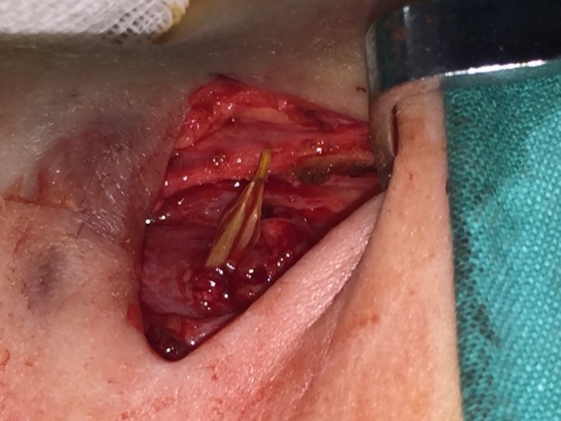
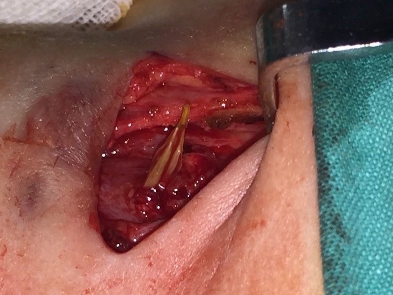
As a result of absence of foreign body in CT scan continuation of medical treatment was offered. Abscess drainage performed at the outpatient clinic and patient was discharged after swellings regression. Because of development of swelling again within a few days, the patient presented to the ENT clinic and surgical intervention was planned (Figure 2).
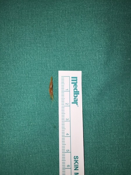
We arrived to the conclusion that the foreign body reached extra laryngeal tissues by penetrating ventricule base. In the postoperative period, dramatic improvement was observed in the patient's clinic. Family who was questioned about foreign body aspiration previously is re-interviewed after surgery. They said that a few days before development of neck mass, they saw him trying to eat grass. DiscussionForeign body aspiration is a common cause of morbidity and mortality in children, especially in children 3-4 years of age [3]. Foreign body existence in oral cavity, oropharynx and larynx is frequently encountered in emergency department and otorhinolaryngologic outpatient clinics. Ingested foreign bodies include common household items such as coins, little toys, marbles, batteries, erasers, rubbers etc. Due to different cultural and eating habits, ingested foreign bodies shows difference in between East and West populations [1]. In China, where seafood nourishment is prevalent, according to Wai Pak et al., who recently described 311 cases of foreign body ingestion in children, most frequently ingested foreign body was fish bones. That was followed by chicken bones, coins, plastic toys and glass fragments [4]. In our country, a tradition; attaching blue beads which are believed to be a protection from evil eyes with a safety pin, has caused safety pin to be an oftenly seen foreign body compared to other countries series [5]. However, coins are reported to be the most common object ingested by children, accounting for up to 70% cases of pediatric foreign body ingestion [1]. Foreign bodies mostly cause acute symptoms, pysicians and families can observe this situation easily [6].However, the foreign body may remain in the food passage due to the fact that the symptoms are not recognized because of low severity or because the swallowed body can not be removed [7]. 80-90% of the foreign bodies that are ingested spontaneously exit, 10-20% require a non-surgical approach; 1% requires surgery [8].With the exception of children under 1 year of age, foreign bodies are implanted less frequently (2-12%) in larynx, but the most dangerous consequences are caused by foreign bodies in this region. If the larynx is fully obstructed, respiratory distress, cyanosis and even respiratory arrest followed by death can occur.Partial obstruction may cause stridor, hoarseness, cough, croup, and dyspnea. Foreign bodies that are thin and sharp (such as fishbone-chicken bone), round and soft (such as grapes, olives) and light and thin (such as egg shells) have higher probability to get stucked in larynx [2].Ingested sharp foreign bodies become lodged in the base of tongue, palatine tonsils, vallecula, pyriform sinus or esophagus [8]. In only a few of them, the foreign body can be periluminally buried into the tissues by penetrating the mucosa, migrating to neighboring tissues and following this unusual path can penetrate vascular structures and visceral organs [7]. Johari et al. reported 3 cases with life-threatening complications after a ingested fishbones migration. The common carotid artery was perforated along its length in one of the cases and a thyroid abscess occured on one and a parapharyngeal abscess occured on the other one. All three cases were adult and successfully treated after neck exploration [9].Chung et al. reported rare complications like deep neck infection, fascial arterial penetration, hematoma at the base of the mouth, and retropharyngeal abscess after ingestion of the fishbone [6].In addition, there are many cases of neck mass after ingestion of fishbone on adults in literature[1]. Although foreign body ingestion is very commonly seen in childhood and adolescence, their penetration from the entrance and migration to neck and is a very rare complication[10]. Pignataro et. al reported in a 15 years old patient with recurrent neck abscess unresponsible to intravenous antibiotics and during surgical drainage found a vegetable fibre. When patients detailed history was being taken patient remembered that a month ago while he was playing with a herb piece in his mouth he felt a sudden pain and his symptoms started after this fact[10]. Landis et al extracted a sharp margined grass blade from an abscess cavity during drainage in a four year old patient with recurrent neck abscess. As the patient's history detailed, family told that a few days before the development of symptoms, they have witnessed the patient was chewing a grass [11]. In our study, as in the two studies above, the fact that the ingested grass ignored by the family and was not mentioned in the story delayed the diagnosis. Radiopacity of ingested foreign bodies help to determine the kind and localization. In the evaluation of radiolucent foreign bodies, it is important to make the necessary examinations by suspicion of family. Especially in children with unexplained findings, the ingestion of a foreign body must always be kept in mind [12]. A patient with the suspicion of foreign body ingestion; positive radiological findings strengthen the diagnosis while negative findings do not exclude the diagnosis in any condition. Failing to take the necessary diagnostic steps based on negative radiological findings can cause serious delays in diagnosis and patients to be followed or hospitalized with wrong diagnosis and treatment for long time [13]. In this case we presented the fact that the foreign body diagnosis was excluded by the CT, which we believed to be reliable, and family disregarding the child's play with grass, distracted us from the possibility of foreign body related complications and delayed diagnosis and treatment. Conclusion Although there are studies reporting foreign body migration to neck in adults, this situation is very rare in kids. While migration of foreign body to deep neck tissues via mucosa penetration and cause to abscess or mass is a rare complication for both adult and childhood patient groups, there are only a few puplications reporting this complication in infants. Foreign bodies that are extracted in those studies are fishbone, chicken bone, chaff, plastic piece, metal wire piece, herb stalk and grass blade. Although a grass blade is not hard and sharp, it may show sharpness depending on the angle during swallowing and may be penetrate tissues. Grass blade ingestion can be ignored or disvalued by both the family and the physician, may not be identified with imaging methods. As in our case it should be kept in mind that harmless looking herb might cause deep neck infection requiring surgery and resistant to medical treatment. Although this case has been evaluated in an experienced clinic for foreign body ingestion and aspiration, not being confronted with this kind of case previously detained diagnosis. We think that presenting that kind of cases into literature is important due to attract physicians attention. References
|
|||||||||
| Keywords : boyun apsesi , migrasyon , yabancı cisim | |||||||||
|