

Authors
|
|||||||
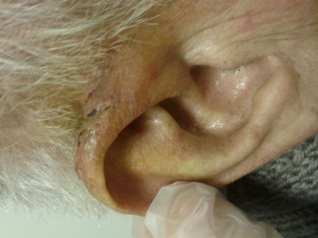
AbstractOtophyma is a rare condition that can present as a stage of rosacea. Rhinophyma lesions can mask the existence of coexisting occult skin cancers, and many types of tumors can mimic a rhinophyma. Although several coexistent malignant tumors have been reported in patients with rhinophyma. However, in the literature there are no reports for otophyma coexisting with any malignant tumor. We report here a case of Squamous Cell Carcinoma(SCC) in a 65-year-old man who presented with a two-month-history of a painful, erythematous lesion on the auricle; dermatological examination suggested an otophyma, and an ENT consultation was requested. We considered the lesion mimicked an otophyma. Histopathological examination revealed an acantholytic SCC. He was treated with surgical excision.IntroductionRosacea is a common, chronic disorder of unknown etiology, which mostly affects middle-aged individuals. In 2002, the National Rosacea Society Expert Committee developed a classification system for rosacea in order to standardize subtypes and variants. Rhinophyma involving the nose is by far the most common type of phymatous growth, whereas other, more rare forms include otophyma (ear), gnatophyma (chin), gnathophyma (jaw), and blepharophyma (eyelids) [1]. With regard to squamous cell carcinomas (SCC), 87% occur in the head and neck regions with 18% of those occurring on the auricle [2]. Squamous cell carcinoma is the second most common type of skin cancer occurring on the external ear [3]. It is a disease found almost exclusively in white men between 60-70 years of age with a history of prolonged sun exposure, and most typically with outdoor occupations [4]. In this study, we present a case with a lesion of the auricle that resembled an otophyma in the initial examination, but the result of diagnostic biopsy revealed SCC. Otophyma has been rarely reported in the literature, and a case with SCC that mimicked otophyma has not been found in our review of the literature. Case ReportA 65-year old male presented with a two month history of an erythematous plaque on the posterosuperior localization of ear. He was investigated at the Başkent University both of Otorhinolaryngology and Dermatology clinics together. He was also a hemodialysis patient because of kidney failure. The patient complained of pain and pruritus. He had central facial erythema (most prominently on the cheeks) with telangiectasias. Our examination revealed that the lesion with plenty of comedones on the erythematous ground without ulceration. It was posterosuperior localization of auricula which was limited posteriorly to the postauricular sulcus . It was resembling an otophyma with inspection (Figure 1).
We performed a biopsy that included the normal skin margins. Histopathological analysis of the biopsy specimen revealed a malignant neoplasia of epithelial origin, characterized by invasive proliferation into the dermis of neoplastic epithelial cell islands. These cells exhibited large nuclei with nuclear pleomorphism, nuclear hyperchromatism, individual cell keratinization, and atypical mitosis.(Figure 2)
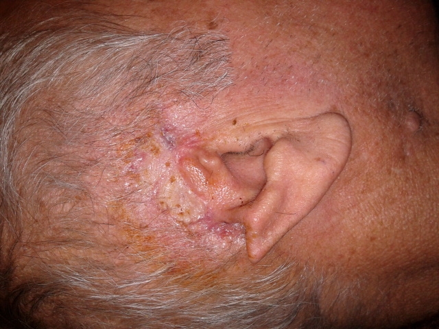
The lesion showed subcutaneous invasion without any ulceration on the skin; palpation revealed a subcutaneous mass of 1 cm size near the helix crura anteriorly, besides on the auricle in posterosuperior localization, a subcutaneous and extensively necrotic lesion. There was moderate cervical lymphadenopathy in the parotid area while the remainder of his examination was relatively benign. There was no temporal bone invasion. We extracted the tumor by wide excision with a surgical margin of 1cm.(Figure 3)
In the analysis of an intraoperative frozen section, a surgical margin was reported to be tumor-negative, and the tissue defect was closed using a split-thickness graft obtained from the thigh. Additional tissue specimens extracted during the reconstruction from the inferior and anterior surgical margins, were referred for pathological analysis. Tissues extracted for the reconstruction were reported to be tumor-positive, and the pathological results revealed a skip metastasis of the tumor that did not show deep invasion. The patient underwent a second surgery, and the surgical limits were enlarged. In the tissues extracted during the second intervention the tumor was indicated to be negative. Pathological investigation of the lesion revealed an acantholytic type squamous cell carcinoma on the grounds of otophyma. Postoperative radiotherapy was not administered due to the result of the oncology consultation. At postop Eighth month a recurrence was observed in the occipital region. After this, PETCT was applied to the patient. PETCT revealed a suspicious mass in the lung requiring biopsy. Patient didn’t accept any surgical treatment and referred to oncology department. DiscussionVarious malignancies, including the basal cell carcinoma [5], angiosarcoma [6], SCC [7,8], and sebaceous carcinoma [9], have been reported to coexist with rhinophyma or to resemble it. Furthermore, it has been suggested that the incidence of certain malignancies is markedly increased in patients with rhinophyma, when compared with those without rhinophyma [10]. But, the described cases of long-standing rhinophyma degenerating into malignancy are still not enough to label rhinophyma as a certain precancerous lesion [11]. The diagnosis of rhinophyma is often straightforward and principally based on the clinical features. However, several clinical characteristics including drainage, ulceration and a rapid growth pattern may be indicative of a malignancy [12]. There are several articles in the literature that report SCC mimicking rhinophyma [13,14], but this is the first study that indicates a SCC mimicking otophyma. In the clinical cases reported to have SCC with rhinophyma, the evolution of rhinophyma was generally long lasting, usually three to 20 years before the observation of a rapid growth rate, and changes in appearance with the development of pain and ulceration only in the few weeks that preceded the diagnosis of a SCC [11]. In our patient, the evolution was too rapid (two months), indicating that the lesion could not be a otophyma. With regard to squamous cell carcinomas, 87% occur in the head and neck regions and 18% of those occur on the auricle. These malignancies occur with the greatest frequency on the superior or posterior portions of the helical rim [15]. In our case, the lesion existed on the same area, because sun exposure plays important role in the development of auricular SCC. Incidences of metastases from SCC of the external ear occur in 5%-18% of the cases, compared with an incidence of metastatic disease in only 0.5%-2 % of the cutaneous SCCs that occur elsewhere [3]. Our case did not occur with lymph node invasion. Involvement of the parotid gland, and cartilage or bone invasion were not investigated. By way of conclusion, lesions that do not show recovery for a long period basically have to be investigated histopathologically, and malignancy has to be excluded. In addition to inspection, the lesion has to be palpated. Lesions appearing as benign by inspection may exist with subcutaneous invasion. References
|
|||||||
| Keywords : Otofima , Skuamöz hücreli karsinom , Rozesea | |||||||


|