

|
|||||||
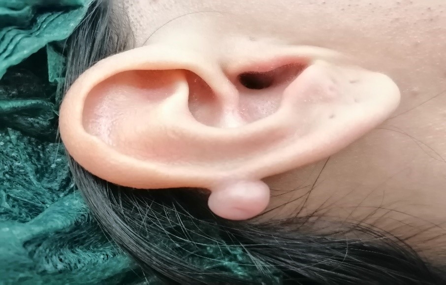
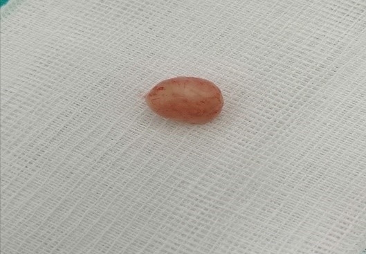
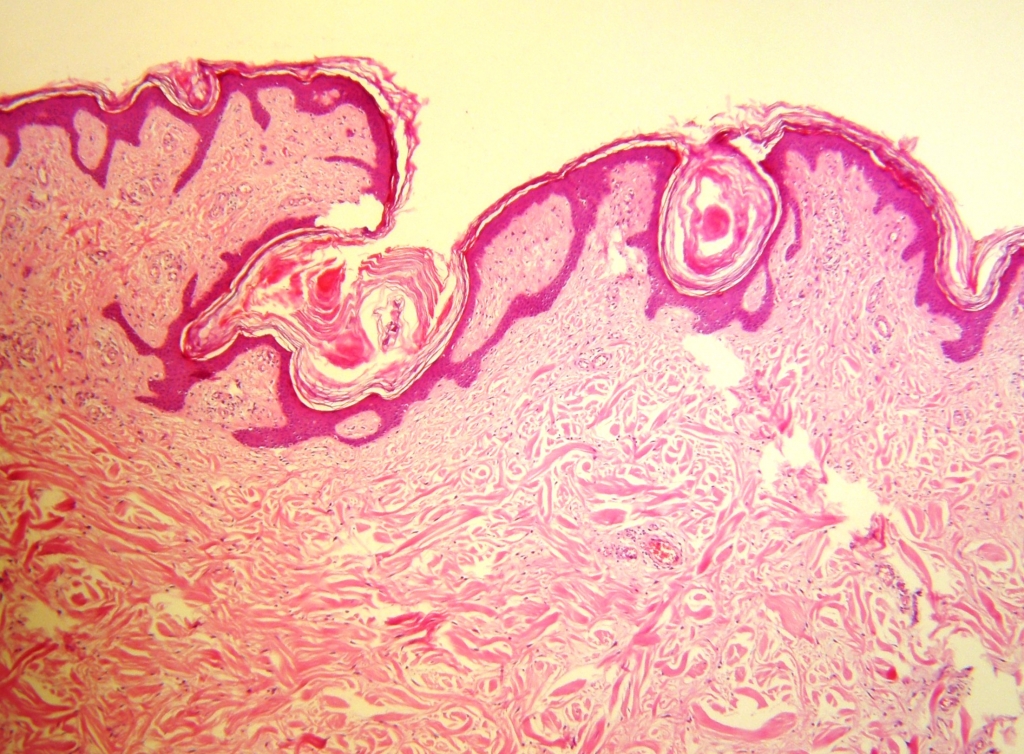
AbstractPiercing is a type of jewelry performed by piercing the skin, underlying fat layer or cartilage.As the use of body piercings became widespread, the number of complications caused by infections, injuries and jewelry has also increased.The aim of this case report was to reveal the clinical features and treatment of our patient who had a mass in the ear with a related piercing history. A 20-year-old female patient, applied to us with her complaints, which started with redness and swelling in the upper part of the right auricle, about 1 year ago, but then gradually grew. On physical examination, there was a painless mass, 0.7x0.5 cm in size in the upper helix of the right auricle, with a smooth surface, slightly darker than the skin, and a minimally eroded area attached to the piercing. The patient had a history of frequent change and use of piercings to the superior helix of the right auricle where the mass was found. An excisional biopsy was performed under local anesthesia. Pathological findings were reported as benign fibroepithelial polyps (FEP). According to our English literature research, no FEP manifested in the auricle and other parts of the body associated with piercing has been reported so far.IntroductionBody piercing is a popular method of body ornamentation, and it is performed with a large needle to make a hole in the skin for jewelry. Modern piercing originated in the USA in the 1970s. For piercing, the auricular cartilage (superior helix), lips, tongue, nose, eyebrow, navel, nipples and genitals are used places. Piercings are associated with poor healing and serious infections due to the avascular nature of the ear cartilage. Approximately 20% of patients have minor complications associated with piercing in the auricula and serious complications are seen in 3% [1]. The earlobe (the soft area under the ear) is the most commonly punctured area and most of the time no complication is observed in this area. Pseudolymphoma [2], lymphocytoma cutis [3], pyogenic granuloma [4], keloid [5] formations are found to be associated with the use of piercing in the auricle. FEP are rare benign mesodermal tumors and they are infrequently observed in our otorhinolaryngology practice. FEPs are also called acrochordons. If it is large, it is called a soft fibroma or pedunculated lipofibroma. FEP is a lesion with a low incidence of malignancy and its etiology is not widely known [6]. FEPs are very rare benign tumors of the auricle. Within the scope of our literature research, we present the first case of FEP associated with piercing in the auricular helix. Case ReportA 20-year-old female patient presented to us with the complaint of a mass in the upper part of the ear that has been developing for about 1 year. The patient stated that she started using piercing 3 years ago and changed piercings frequently in last 3 years. About 2 years later, she started having with mild redness and swelling which did not worry her at first, but she worried about the swelling when it became consistent and began to growing in recent months. And she applied to our clinic. She declared that she had stopped using her piercing few months prior to her admittance. She stated that she mostly used piercings immitated. The piercing she used did not contain gold or silver. Ear nose throat examination was normal. There was no lymphadenopathy in the neck. She did not have any systemic disease or concomitant allergic disease and she declared no continuous medication use. On physical examination, there were a total of 4 places of piercing entry in the right auricle. In the upper part of the helix of the right auricle, there was a 0.7x0.5 cm sized, slightly darker, smooth, painless mass with a eroded surface that did not bleed by touching to the attachment site. The patient was informed about the medical procedure . A local anesthetic was injected into the auricular skin and a small superficial incision was made to safely remove the poly. The mass was excised from the intact tissue margin under local anesthesia. histopathological examination was reported ad fibroepithelial polyp. We presented the surgical treatment of this benign mass and there were no early postoperative complications.
The patient was discharged on the same day. Sutures were removed in the first week after the operation. Complete recovery was seen at the end of the second week. DiscussionIn our case, there was overuse of the auricle suggesting the presence of mechanical stimulation of the superior helix of the auricle. Therefore, this is the first case report of FEP related to the use of piercing of the auricula. To date, ear-related FEPs have been reported in the outer ear canal [7] and middle ear [8]. However, piercing-associated auricular FEP has not been reported. Several theories about the formation of polyps have been proposed so far. The first of these is the irreversible obstruction of lymphatic ducts with congestion as a result of chronic infections [9]. Another theory is that FEP occurs after chronic irritation [10]. In our case, chronic irritation was also present. But there was no clinical finding suggesting previous history of aural polyp or other ear disorders. Therefore, we strongly believe that formation mechanism of our patient's polyp was mechanical stimulation as chronic irritation and repetitive injury of the auricle caused by piercing use. Possibly, contributing pathogenesis by leading to release of signalling molecules such as, cytokines and growth factors which are the main regulators of metabolic events, promoters of tumoural formations and inflammatory processes like stromal edema, chronic inflammatory infiltrations as they are observed in fibroepithelial polyps [11]. We also think that repetitive perforation injury followed by an increased blood flow enhanced the inflammation and fibrotic events. Although FEPs are benign tumors that grow slowly, they require surgical excision due to metaplasia or malignant transformation risk.[9] We also performed excision of the tumor in our case. There are a few important points that we would like to emphasize in our case. People should be more careful while using piercing and should be more aware about the complications of piercing. As in our case, piercing is used for aesthetic purposes and it can cause FEP as a result of chronic irritation. Although these tumors are benign tumors, they can be percieved as a malignant tumor by patients and cause anxiety. Here, this benign tumor was treated with surgery by considering the patient's current symptoms. In this study, we reported a case of piercing-associated FEP of the superior auricular helix. FEP should be considered in the differential diagnosis of auricular piercing problems and should be excised since there is a low risk of transformation to a malignancy. References
|
|||||||
| Keywords : Dermatopatoloji , irritan dermatit , patogenez , yaşam kalitesi | |||||||

|